

The effect of tumor-targeted photodynamic therapy (PDT) was improved by designing nanotheranostics to promote oxygenation in a tumor microenvironment (TME) wherein hypoxia, acidosis, and the elevated levels of H2O2 are three main characteristics. In this study, a carbon dot (CD) PDT agent recently developed by our group was firstly applied as reducing agent to react with potassium permanganate for fabricating CDs/manganese dioxide (CDs/MnO2) composites, which were in turn modified with polyethylene glycol (PEG) to form water-soluble CDs/MnO2-PEG nanohybrids. In a normal physiological environment, the as-prepared nanohybrids exhibited quenched fluorescence, weak singlet oxygen generation, and low magnetic resonance imaging (MRI) signal. However, given the high sensitivity of MnO2 to the TME, the CDs/MnO2-PEG nanohybrids changed from an “off” to an “on” state with synchronously enhanced fluorescence, singlet oxygen generation, and MRI signal in the TME.
and the Strategic Priority Research Program of the Chinese Academy of Sciences(XDB17000000)
the National Natural Science Foundation of China(51472252)
This work was supported by the National Natural Science Foundation of China (51472252 and 51572269), and the Strategic Priority Research Program of the Chinese Academy of Sciences (XDB17000000).
The authors declare that they have no conflict of interest.
Wang P and Ge J supervised the project. Chen S designed and carried out the experiments, analyzed the data and wrote the manuscript. Jia Q, Zheng X and Wen Y helped with the synthesis of the CDs/MnO2-PEG. Liu W and Zhang H helped with the photodynamic therapy.
Supplementary information
Supporting data are available in the online version of the paper.
[1] Kunjachan S, Ehling J, Storm G, et al. Noninvasive imaging of nanomedicines and nanotheranostics: principles, progress, and prospects. Chem Rev, 2015, 115: 10907-10937 CrossRef PubMed Google Scholar
[2] Sun Q, Sun X, Ma X, et al. Integration of nanoassembly functions for an effective delivery cascade for cancer drugs. Adv Mater, 2014, 26: 7615-7621 CrossRef PubMed Google Scholar
[3] Singh RK, Patel KD, Leong KW, et al. Progress in nanotheranostics based on mesoporous silica nanomaterial platforms. ACS Appl Mater Interfaces, 2017, 9: 10309-10337 CrossRef Google Scholar
[4] Huang H, Lovell JF. Advanced functional nanomaterials for theranostics. Adv Funct Mater, 2017, 27: 1603524 CrossRef PubMed Google Scholar
[5] Huo M, Wang L, Chen Y, et al. Tumor-selective catalytic nanomedicine by nanocatalyst delivery. Nat Commun, 2017, 8: 357 CrossRef PubMed Google Scholar
[6] Shi J, Kantoff PW, Wooster R, et al. Cancer nanomedicine: progress, challenges and opportunities. Nat Rev Cancer, 2017, 17: 20-37 CrossRef PubMed Google Scholar
[7] Anchordoquy TJ, Barenholz Y, Boraschi D, et al. Mechanisms and barriers in cancer nanomedicine: addressing challenges, looking for solutions. ACS Nano, 2017, 11: 12-18 CrossRef Google Scholar
[8] Ma Y, Li X, Li A, et al. H2S-activable MOF nanoparticle photosensitizer for effective photodynamic therapy against cancer with controllable singlet-oxygen release. Angew Chem Int Ed, 2017, 56: 13752-13756 CrossRef PubMed Google Scholar
[9] Hu D, Sheng Z, Gao G, et al. Activatable albumin-photosensitizer nanoassemblies for triple-modal imaging and thermal-modulated photodynamic therapy of cancer. Biomaterials, 2016, 93: 10-19 CrossRef PubMed Google Scholar
[10] Luo D, Carter KA, Razi A, et al. Doxorubicin encapsulated in stealth liposomes conferred with light-triggered drug release. Biomaterials, 2016, 75: 193-202 CrossRef PubMed Google Scholar
[11] Lucky SS, Soo KC, Zhang Y. Nanoparticles in photodynamic therapy. Chem Rev, 2015, 115: 1990-2042 CrossRef PubMed Google Scholar
[12] Yuan Y, Zhang CJ, Gao M, et al. Specific light-up bioprobe with aggregation-induced emission and activatable photoactivity for the targeted and image-guided photodynamic ablation of cancer cells. Angew Chem Int Ed, 2015, 54: 1780-1786 CrossRef PubMed Google Scholar
[13] Guo L, Liu W, Niu G, et al. Polymer nanoparticles with high photothermal conversion efficiency as robust photoacoustic and thermal theranostics. J Mater Chem B, 2017, 5: 2832-2839 CrossRef Google Scholar
[14] Xing R, Liu K, Jiao T, et al. An injectable self-assembling collagen-gold hybrid hydrogel for combinatorial antitumor photothermal/photodynamic therapy. Adv Mater, 2016, 28: 3669-3676 CrossRef PubMed Google Scholar
[15] Jia Q, Chen M, Liu Q, et al. Ethylene glycol-mediated synthetic route for production of luminescent silicon nanorod as photodynamic therapy agent. Sci China Mater, 2017, 60: 881-891 CrossRef Google Scholar
[16] Wen R, Lv X, Yang T, et al. Albumin nanoreactor-templated synthesis of Gd2O3/CuS hybrid nanodots for cancer theranostics. Sci China Mater, 2017, 60: 554-562 CrossRef Google Scholar
[17] Yu Z, Wang M, Pan W, et al. Tumor microenvironment-triggered fabrication of gold nanomachines for tumor-specific photoacoustic imaging and photothermal therapy. Chem Sci, 2017, 8: 4896-4903 CrossRef PubMed Google Scholar
[18] Kanamala M, Wilson WR, Yang M, et al. Mechanisms and biomaterials in pH-responsive tumour targeted drug delivery: a review. Biomaterials, 2016, 85: 152-167 CrossRef PubMed Google Scholar
[19] Mura S, Nicolas J, Couvreur P. Stimuli-responsive nanocarriers for drug delivery. Nat Mater, 2013, 12: 991-1003 CrossRef PubMed ADS Google Scholar
[20]
Mou
J,
Chen
Y,
Ma
M, et al.
Facile synthesis of liposome/Cu2?
[21]
Mathiyazhakan
M,
Upputuri
PK,
Sivasubramanian
K, et al.
[22] Dai Y, Xu C, Sun X, et al. Nanoparticle design strategies for enhanced anticancer therapy by exploiting the tumour microenvironment. Chem Soc Rev, 2017, 46: 3830-3852 CrossRef PubMed Google Scholar
[23] Khawar IA, Kim JH, Kuh HJ. Improving drug delivery to solid tumors: Priming the tumor microenvironment. J Control Release, 2015, 201: 78-89 CrossRef PubMed Google Scholar
[24] Wu T, Dai Y. Tumor microenvironment and therapeutic response. Cancer Lett, 2016, 387: 61-68 CrossRef PubMed Google Scholar
[25] Cheng H, Zhu JY, Li SY, et al. An O2 self-sufficient biomimetic nanoplatform for highly specific and efficient photodynamic therapy. Adv Funct Mater, 2016, 26: 7847-7860 CrossRef Google Scholar
[26] Chen H, Tian J, He W, et al. H2O2-activatable and O2-evolving nanoparticles for highly efficient and selective photodynamic therapy against hypoxic tumor cells. J Am Chem Soc, 2015, 137: 1539-1547 CrossRef PubMed Google Scholar
[27] Chen Q, Feng L, Liu J, et al. Intelligent albumin-MnO2 nanoparticles as pH-/H2O2-responsive dissociable nanocarriers to modulate tumor hypoxia for effective combination therapy. Adv Mater, 2016, 28: 7129-7136 CrossRef PubMed Google Scholar
[28] Yang G, Zhang R, Liang C, et al. Manganese dioxide coated WS2@Fe3O4/sSiO2 nanocomposites for pH-responsive MR imaging and oxygen-elevated synergetic therapy. Small, 2018, 14: 1702664 CrossRef PubMed Google Scholar
[29] Liu J, Chen Q, Zhu W, et al. Nanoscale-coordination-polymer-shelled manganese dioxide composite nanoparticles: a multistage redox/pH/H2O2-responsive cancer theranostic nanoplatform. Adv Funct Mater, 2017, 27: 1605926 CrossRef Google Scholar
[30] Yuan F, Li S, Fan Z, et al. Shining carbon dots: Synthesis and biomedical and optoelectronic applications. Nano Today, 2016, 11: 565-586 CrossRef Google Scholar
[31] Lim SY, Shen W, Gao Z. Carbon quantum dots and their applications. Chem Soc Rev, 2015, 44: 362-381 CrossRef PubMed Google Scholar
[32] Zhu S, Meng Q, Wang L, et al. Highly photoluminescent carbon dots for multicolor patterning, sensors, and bioimaging. Angew Chem Int Ed, 2013, 52: 3953-3957 CrossRef PubMed Google Scholar
[33] Chen Y, Shi J. Mesoporous carbon biomaterials. Sci China Mater, 2015, 58: 241-257 CrossRef Google Scholar
[34] Roy E, Patra S, Madhuri R, et al. Carbon dot/TAT peptide co-conjugated bubble nanoliposome for multicolor cell imaging, nuclear-targeted delivery, and chemo/photothermal synergistic therapy. Chem Eng J, 2017, 312: 144-157 CrossRef Google Scholar
[35] Huang Q, Lin X, Zhu JJ, et al. Pd-Au@carbon dots nanocomposite: Facile synthesis and application as an ultrasensitive electrochemical biosensor for determination of colitoxin DNA in human serum. Biosens Bioelectron, 2017, 94: 507-512 CrossRef PubMed Google Scholar
[36]
Gao
N,
Yang
W,
Nie
H, et al.
Turn-on theranostic fluorescent nanoprobe by electrostatic self-assembly of carbon dots with doxorubicin for targeted cancer cell imaging,
[37] Ge J, Lan M, Liu W, et al. Graphene quantum dots as efficient, metal-free, visible-light-active photocatalysts. Sci China Mater, 2016, 59: 12-19 CrossRef Google Scholar
[38] Wang Z, Yuan F, Li X, et al. 53% Efficient red emissive carbon quantum dots for high color rendering and stable warm white-light-emitting diodes. Adv Mater, 2017, 29: 1702910 CrossRef PubMed Google Scholar
[39] Ong WJ, Putri LK, Tan YC, et al. Unravelling charge carrier dynamics in protonated g-C3N4 interfaced with carbon nanodots as co-catalysts toward enhanced photocatalytic CO2 reduction: A combined experimental and first-principles DFT study. Nano Res, 2017, 10: 1673-1696 CrossRef Google Scholar
[40] Huang P, Lin J, Wang X, et al. Light-triggered theranostics based on photosensitizer-conjugated carbon dots for simultaneous enhanced-fluorescence imaging and photodynamic therapy. Adv Mater, 2012, 24: 5104-5110 CrossRef PubMed Google Scholar
[41]
Choi
Y,
Kim
S,
Choi
MH, et al.
Highly biocompatible carbon nanodots for simultaneous bioimaging and targeted photodynamic therapy
[42] Zheng M, Liu S, Li J, et al. Integrating oxaliplatin with highly luminescent carbon dots: an unprecedented theranostic agent for personalized medicine. Adv Mater, 2014, 26: 3554-3560 CrossRef PubMed Google Scholar
[43] Karthik S, Saha B, Ghosh SK, et al. Photoresponsive quinoline tethered fluorescent carbon dots for regulated anticancer drug delivery. Chem Commun, 2013, 49: 10471-10473 CrossRef PubMed Google Scholar
[44] Zhang M, Wang W, Zhou N, et al. Near-infrared light triggered photo-therapy, in combination with chemotherapy using magnetofluorescent carbon quantum dots for effective cancer treating. Carbon, 2017, 118: 752-764 CrossRef Google Scholar
[45] Fan Z, Zhou S, Garcia C, et al. pH-responsive fluorescent graphene quantum dots for fluorescence-guided cancer surgery and diagnosis. Nanoscale, 2017, 9: 4928-4933 CrossRef PubMed Google Scholar
[46]
Zheng
DW,
Li
B,
Li
CX, et al.
Carbon-dot-decorated carbon nitride nanoparticles for enhanced photodynamic therapy against hypoxic tumor
[47]
Zeng
Q,
Shao
D,
He
X, et al.
Carbon dots as a trackable drug delivery carrier for localized cancer therapy
[48]
Feng
T,
Ai
X,
An
G, et al.
Charge-convertible carbon dots for imaging-guided drug delivery with enhanced
[49] Ge J, Lan M, Zhou B, et al. A graphene quantum dot photodynamic therapy agent with high singlet oxygen generation. Nat Commun, 2014, 5: 4596 CrossRef PubMed ADS Google Scholar
[50]
Ge
J,
Jia
Q,
Liu
W, et al.
Carbon dots with intrinsic theranostic properties for bioimaging, red-light-triggered photodynamic/photothermal simultaneous therapy
[51] Ge J, Jia Q, Liu W, et al. Red-emissive carbon dots for fluorescent, photoacoustic, and thermal theranostics in living mice. Adv Mater, 2015, 27: 4169-4177 CrossRef PubMed Google Scholar
[52] Cai QY, Li J, Ge J, et al. A rapid fluorescence “switch-on” assay for glutathione detection by using carbon dots–MnO2 nanocomposites. Biosens Bioelectron, 2015, 72: 31-36 CrossRef PubMed Google Scholar
[53] Zhu W, Dong Z, Fu T, et al. Modulation of hypoxia in solid tumor microenvironment with MnO2 nanoparticles to enhance photodynamic therapy. Adv Funct Mater, 2016, 26: 5490-5498 CrossRef Google Scholar
[54] Chen Y, Ye D, Wu M, et al. Break-up of two-dimensional MnO2 nanosheets promotes ultrasensitive pH-triggered theranostics of cancer. Adv Mater, 2014, 26: 7019-7026 CrossRef PubMed Google Scholar
[55] Prasad P, Gordijo CR, Abbasi AZ, et al. Correction to multifunctional albumin–MnO2 nanoparticles modulate solid tumor microenvironment by attenuating hypoxia, acidosis, vascular endothelial growth factor and enhance radiation response. ACS Nano, 2014, 8: 6510-6510 CrossRef Google Scholar
[56] Abbasi AZ, Gordijo CR, Amini MA, et al. Hybrid manganese dioxide nanoparticles potentiate radiation therapy by modulating tumor hypoxia. Cancer Res, 2016, 76: 6643-6656 CrossRef PubMed Google Scholar
[57] Song M, Liu T, Shi C, et al. Bioconjugated manganese dioxide nanoparticles enhance chemotherapy response by priming tumor-associated macrophages toward m1-like phenotype and attenuating tumor hypoxia. ACS Nano, 2016, 10: 633-647 CrossRef Google Scholar
[58] Fan W, Bu W, Shen B, et al. Intelligent MnO2 nanosheets anchored with upconversion nanoprobes for concurrent pH-/H2O2-responsive UCL imaging and oxygen-elevated synergetic therapy. Adv Mater, 2015, 27: 4155-4161 CrossRef PubMed Google Scholar
[59] Yang G, Xu L, Chao Y, et al. Hollow MnO2 as a tumor-microenvironment-responsive biodegradable nano-platform for combination therapy favoring antitumor immune responses. Nat Commun, 2017, 8: 902 CrossRef PubMed Google Scholar
[60] Gao S, Wang G, Qin Z, et al. Oxygen-generating hybrid nanoparticles to enhance fluorescent/photoacoustic/ultrasound imaging guided tumor photodynamic therapy. Biomaterials, 2017, 112: 324-335 CrossRef PubMed Google Scholar
[61] Fang J, Nakamura H, Maeda H. The EPR effect: unique features of tumor blood vessels for drug delivery, factors involved, and limitations and augmentation of the effect. Adv Drug Deliver Rev, 2011, 63: 136-151 CrossRef PubMed Google Scholar
[62] Peng J, Dong M, Ran B, et al. “One-for-all”-type, biodegradable prussian blue/manganese dioxide hybrid nanocrystal for trimodal imaging-guided photothermal therapy and oxygen regulation of breast cancer. ACS Appl Mater Interfaces, 2017, 9: 13875-13886 CrossRef Google Scholar
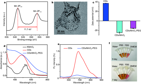
Figure 1
Characterizations of CDs/MnO2-PEG nanohybrids. (a) XPS spectrum of CDs/MnO2 nanohybrids. (b) A TEM image of CDs/MnO2-PEG nanohybrids (scale bar:
Scheme 1
Schematic illustration of CDs/MnO2-PEG nanohybrids as a multimodal theranostics for the MR/FL imaging-guided PDT.
Figure 2
pH/HO2-response of CDs/MnO2-PEG nanohybrids. (a) UV-vis-NIR spectra and (b) FL spectra of CDs/MnO2-PEG nanohybrids at pH 6.5 and 7.4, respectively. (c) Simultaneous O2 generation in acidic H2O2 solutions
Figure 3
Figure 4
Figure 5


Copyright 2019 Science China Press Co., Ltd. 科学大众杂志社有限责任公司 版权所有
京ICP备18024590号-1

 Download PDF


