

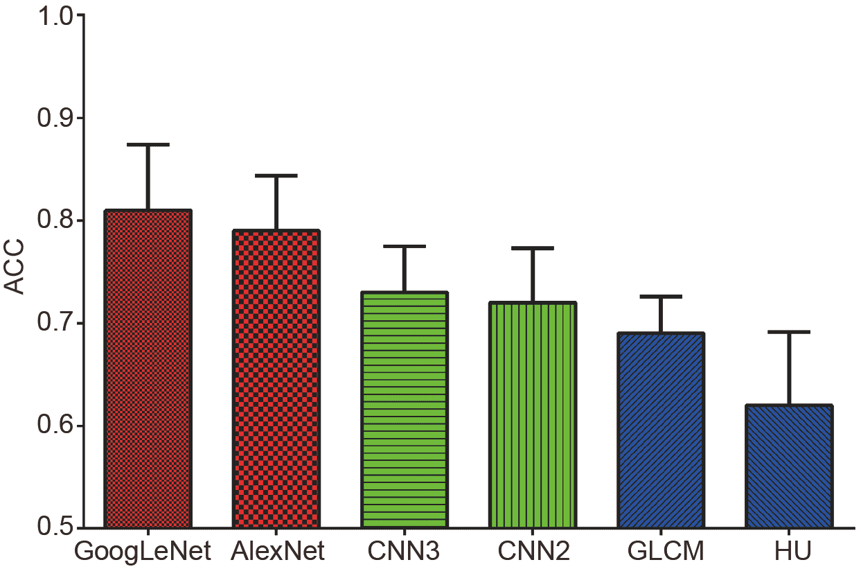
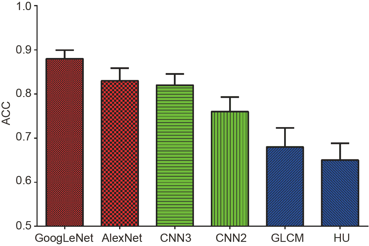
Machine learning can help differentiating benign and malignant lesions seen on mammographic images. Conventional models require handcrafting features for lesion representation. Due to insufficient medical instances, the performance of convolutional neural networks (CNNs) can be further increased. This study makes use of transfer learning for mammographic breast lesion diagnosis and deep neural network (DNN) models pre-trained with large-scale natural images are employed. The diagnosis performance is evaluated with the prediction accuracy (ACC) and the area under the curve (AUC) on average. A histologically verified database is analyzed which contains 406 lesions (230 benign and 176 malignant). Involved models include transferred DNNs (GoogLeNet and AlexNet), shallow CNNs (CNN2 and CNN3) that are fully trained with medical instances and boosted by support vector machine (SVM), and two conventional methods which combine handcrafted features and SVM for lesion diagnosis. Experimental results indicate that GoogLeNet achieves the best performance (ACC=0.81, AUC=0.88), followed by AlexNet (ACC=0.79, AUC=0.83) and CNN3 (ACC=0.73, AUC=0.82). Knowledge transfer can improve the mammographic breast cancer diagnosis, while its wide application still requires further verification in medical imaging domain.
the National Key Research and Develop Program of China(Grant,No.,2016YFC0105102)
the Shenzhen Key Technical Research Project(Grant,No.,JSGG20160229203812944)
the Leading Talent of Special Support Project in Guangdong(Grant,No.,2016TX03R139)
the Science Foundation of Guangdong(Grant,Nos.,2017B020229002,&,2014A030312006)
the National Natural Science Foundation of China(Grant,No.,61471349)
the Major Scientific Research Project for Universities of Guangdong Province(Grant,No.,2016KTSCX167)
The authors thank the Breast Cancer Digital Repository Consortium for sharing the database BCDR-F03. This work was supported in part by the National Key Research and Development Program of China (Grant No. 2016YFC0105102), the Leading Talent of Special Support Project in Guangdong (Grant No. 2016TX03R139), the Shenzhen Key Technical Research Project (Grant No. JSGG20160229203812944), the Science Foundation of Guangdong (Grant Nos. 2017B020229002, 2015B020233011 & 2014A030312006), the National Natural Science Foundation of China (Grant No. 61871374), the Beijing Center for Mathematics and Information Interdisciplinary Sciences, and the Major Scientific Research Project for Universities of Guangdong Province (Grant No. 2016KTSCX167).
[1] DeSantis C E, Ma J, Goding Sauer A, et al. Breast cancer statistics, 2017, racial disparity in mortality by state. CA-Cancer J Clin, 2017, 67: 439-448 CrossRef PubMed Google Scholar
[2] Fan L, Strasser-Weippl K, Li J J, et al. Breast cancer in China. Lancet Oncol, 2014, 15: e279-e289 CrossRef Google Scholar
[3] Mittal S, Kaur H, Gautam N, et al. Biosensors for breast cancer diagnosis: A review of bioreceptors, biotransducers and signal amplification strategies. Biosens Bioelectron, 2017, 88: 217-231 CrossRef PubMed Google Scholar
[4] Bahl M, Barzilay R, Yedidia A B, et al. High-risk breast lesions: A machine learning model to predict pathologic upgrade and reduce unnecessary surgical excision. Radiology, 2018, 286: 810-818 CrossRef PubMed Google Scholar
[5] Tan M, Pu J, Zheng B. Reduction of false-positive recalls using a computerized mammographic image feature analysis scheme. Phys Med Biol, 2014, 59: 4357-4373 CrossRef PubMed ADS Google Scholar
[6] Elter M, Horsch A. CADx of mammographic masses and clustered microcalcifications: A review. Med Phys, 2009, 36: 2052-2068 CrossRef PubMed ADS Google Scholar
[7] Tang J, Rangayyan R M, Xu J, et al. Computer-aided detection and diagnosis of breast cancer with mammography: Recent advances. IEEE Trans Inform Technol Biomed, 2009, 13: 236-251 CrossRef PubMed Google Scholar
[8] Moura D C, Guevara López M A. An evaluation of image descriptors combined with clinical data for breast cancer diagnosis. Int J Comput Ass Rad, 2013, 8: 561-574 CrossRef PubMed Google Scholar
[9] Chang C C, Lin C J. LIBSVM: A library for support vector machines. ACM Trans Intell Syst Technol, 2011, 2: 1-27 CrossRef Google Scholar
[10] Ramos-Pollán R, Guevara-López M A, Suárez-Ortega C, et al. Discovering mammography-based machine learning classifiers for breast cancer diagnosis. J Med Syst, 2012, 36: 2259-2269 CrossRef PubMed Google Scholar
[11] Khan S, Hussain M, Aboalsamh H, et al. A comparison of different Gabor feature extraction approaches for mass classification in mammography. Multimed Tools Appl, 2017, 76: 33-57 CrossRef Google Scholar
[12] Wang Y, Li J, Gao X. Latent feature mining of spatial and marginal characteristics for mammographic mass classification. Neurocomputing, 2015, 144: 107-118 CrossRef Google Scholar
[13] Xie W, Li Y, Ma Y. Breast mass classification in digital mammography based on extreme learning machine. Neurocomputing, 2016, 173: 930-941 CrossRef Google Scholar
[14] Li Y, Chen H, Wei X, et al. Mass classification in mammograms based on two-concentric masks and discriminating texton. Pattern Recognit, 2016, 60: 648-656 CrossRef Google Scholar
[15] Benndorf M, Burnside E S, Herda C, et al. External validation of a publicly available computer assisted diagnostic tool for mammographic mass lesions with two high prevalence research datasets. Med Phys, 2015, 42: 4987-4996 CrossRef PubMed ADS Google Scholar
[16] Hu K, Yang W, Gao X. Microcalcification diagnosis in digital mammography using extreme learning machine based on hidden Markov tree model of dual-tree complex Wavelet transform. Expert Syst Appl, 2017, 86: 135-144 CrossRef Google Scholar
[17] Samala R K, Chan H P, Hadjiiski L M, et al. Multi-task transfer learning deep convolutional neural network: Application to computer-aided diagnosis of breast cancer on mammograms. Phys Med Biol, 2017, 62: 8894. Google Scholar
[18] Tajbakhsh N, Shin J Y, Gurudu S R, et al. Convolutional neural networks for medical image analysis: Full training or fine tuning?. IEEE Trans Med Imag, 2016, 35: 1299-1312 CrossRef PubMed Google Scholar
[19] Shin H C, Roth H R, Gao M, et al. Deep convolutional neural networks for computer-aided detection: CNN architectures, dataset characteristics and transfer learning. IEEE Trans Med Imag, 2016, 35: 1285-1298 CrossRef PubMed Google Scholar
[20] LeCun Y, Bengio Y, Hinton G. Deep learning. Nature, 2015, 521: 436–444. Google Scholar
[21] Arevalo J, González F A, Ramos-Pollán R, et al. Representation learning for mammography mass lesion classification with convolutional neural networks. Comput Methods Programs Biomed, 2016, 127: 248-257 CrossRef PubMed Google Scholar
[22] Yosinski J, Clune J, Bengio Y, et al. How transferable are features in deep neural networks. Adv Neural Inform Process Syst, 2014: 3320–3328. Google Scholar
[23] Carneiro G, Nascimento J, Bradley A P. Unregistered multiview mammogram analysis with pre-trained deep learning models. In: Navab N, Hornegger J, Wells W, et al, Eds. Medical Image Computing and Computer-Assisted Intervention—MICCAI 2015. Lecture Notes in Computer Science, Vol. 9351. Cham: Springer, 2015. 652–660. Google Scholar
[24] Huynh B Q, Li H, Giger M L. Digital mammographic tumor classification using transfer learning from deep convolutional neural networks. J Med Imag, 2016, 3: 034501 CrossRef PubMed Google Scholar
[25] Krizhevsky A, Sutskever I, Hinton G E. ImageNet classification with deep convolutional neural networks. Adv Neural Inform Process Syst, 2012: 1097–1105. Google Scholar
[26] Szegedy C, Liu W, Jia Y, et al. Going deeper with convolutions. In: IEEE conference on Computer Vision and Pattern Recognition. Boston, 2015. 1–9. Google Scholar
[27] Russakovsky O, Deng J, Su H, et al. ImageNet large scale visual recognition challenge. Int J Comput Vis, 2015, 115: 211-252 CrossRef Google Scholar
[28] Otsu N. A threshold selection method from gray-level histograms. Automatica, 1975, 11: 23–27. Google Scholar
[29] Soh L K, Tsatsoulis C. Texture analysis of SAR sea ice imagery using gray level co-occurrence matrices. IEEE Trans Geosci Remote Sens, 1999, 37: 780-795 CrossRef ADS Google Scholar
[30] Hu M K. Visual pattern recognition by moment invariants. IRE Trans Inform Theor, 1962, 8: 179–187. Google Scholar
[31] Jia Y, Shelhamer E, Donahue J, et al. Caffe: Convolutional architecture for fast feature embedding. In: Proceedings of the 22nd ACM international conference on Multimedia. Orlando, 2014. 675–678. Google Scholar
[32] Pan S J, Yang Q. A survey on transfer learning. IEEE Trans Knowl Data Eng, 2010, 22: 1345-1359 CrossRef Google Scholar
[33] Bergstra J, Bengio Y. Random search for hyper-parameter optimization. J Mac Learn Res, 2012, 13: 281–305. Google Scholar
[34] Zhang Z, Dai G, Liang X, et al. Can signal-to-noise ratio perform as a baseline indicator for medical image quality assessment. IEEE Access, 2018, 6: 11534-11543 CrossRef Google Scholar
[35] Casti P, Mencattini A, Salmeri M, et al. Towards localization of malignant sites of asymmetry across bilateral mammograms. Comput Methods Programs Biomed, 2017, 140: 11-18 CrossRef PubMed Google Scholar
[36] He K, Zhang X, Ren S, et al. Deep residual learning for image recognition. In: Proceedings of the IEEE conference on computer vision and pattern recognition. Las Vegas, 2016. 770–778. Google Scholar
[37] Xiao T, Liu L, Li K, et al. Comparison of transferred deep neural networks in ultrasonic breast masses discrimination. Biomed Res Int, 2018, 2018: 1-9 CrossRef PubMed Google Scholar
[38] Yassin N I R, Omran S, El Houby E M F, et al. Machine learning techniques for breast cancer computer aided diagnosis using different image modalities: A systematic review. Comput Methods Programs Biomed, 2018, 156: 25-45 CrossRef PubMed Google Scholar
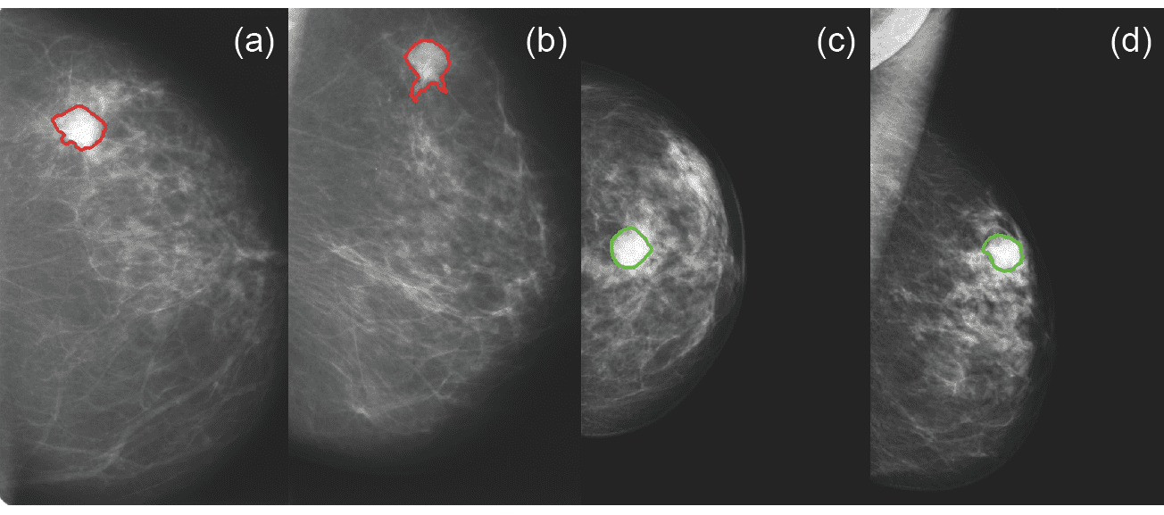
Figure 1
Breast lesion visualization. The malignant lesion and benign ones are shown in craneocaudal ((a), (c)) and mediolateral oblique ((b), (d)) views. The coordinates of the red and green contours are provided in the BCDR-F03.
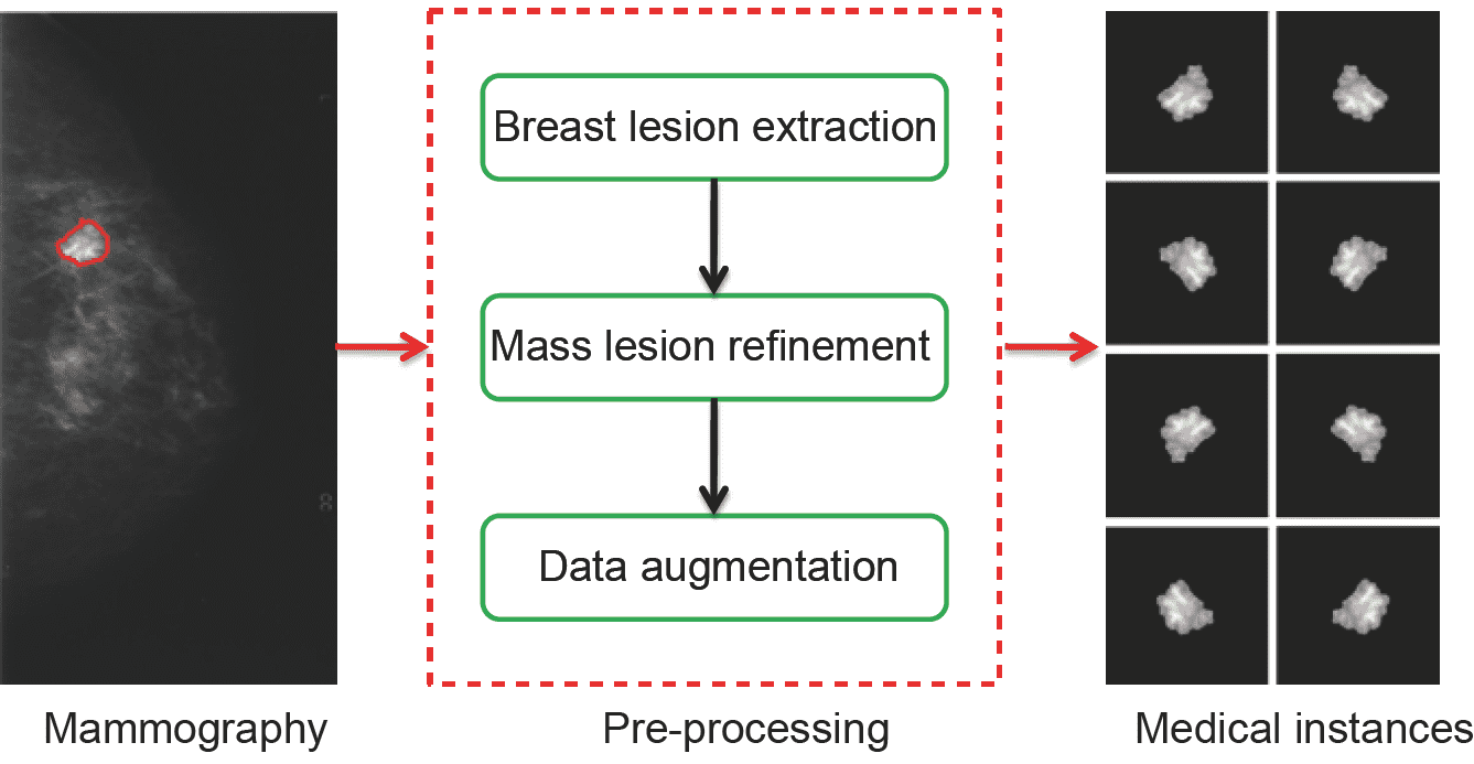
Figure 2
Medical image pre-processing. The diagram consists of breast lesion extraction, mass lesion refinement and data augmentation. After image pre-processing, seven new medical instances are added to each mass.
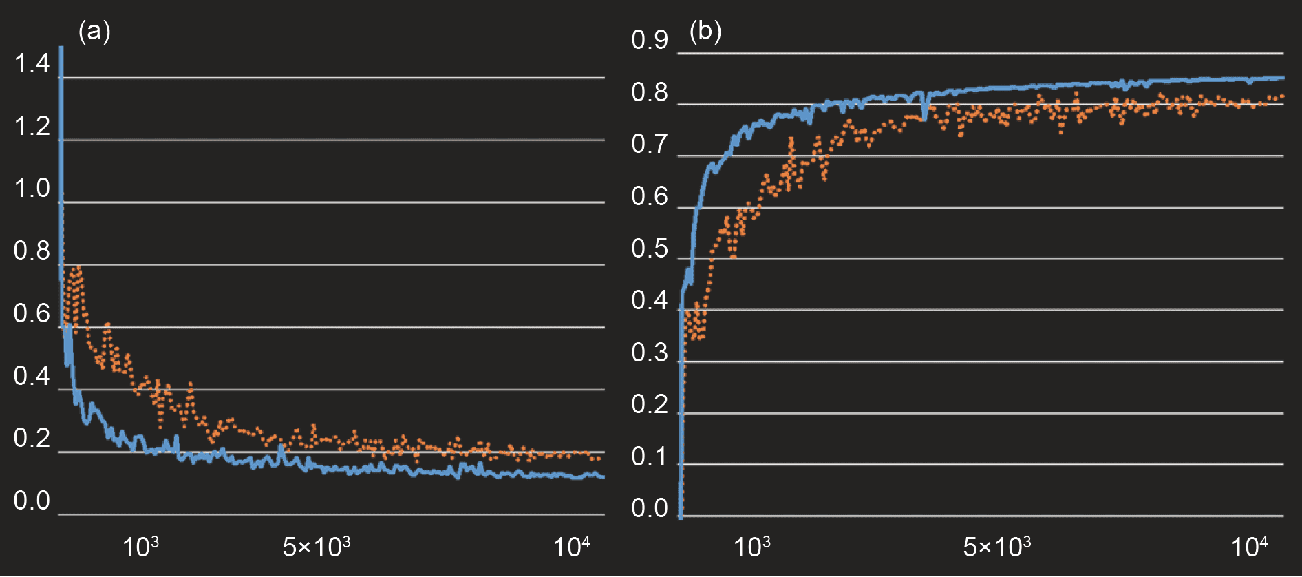
Figure 3
The convergence analysis in model transferring. The brown and the blue dots refer to the convergence procedure when fine-tuning of AlexNet and GoogLeNet, respectively. (a) Loss in training; (b) accuracy in validation.
Figure 4
(Color online)
Figure 5
(Color online)
Age range | Patient cases |
[20, 40) | 28 |
[40, 60) | 215 |
[60, 80) | 130 |
[80, 100) | 14 |
Others | 19 |
View number | Patient cases |
1 | 89 |
2 | 310 |
3 | 5 |
4 | 1 |
8 | 1 |
")

Copyright 2019 Science China Press Co., Ltd. 科学大众杂志社有限责任公司 版权所有
京ICP备18024590号-1

 Download PDF