

Microparticles have a demonstrated value for drug delivery systems.
The attempts to develop this technology focus on the generation of
featured microparticles for improving the function of the systems.
Here, we present a new type of microparticles with gelatin methacrylate
(GelMa) cores and poly(L-lactide-co-glycolide) (PLGA) shells for synergistic
and sustained drug delivery applications. The microparticles were
fabricated by using GelMa aqueous solution and PLGA oil solution as
the raw materials of the microfluidic double emulsion templates, in
which hydrophilic and hydrophobic actives, such as doxorubicin
National Science Foundation of Jiangsu(BK20140028)
Scientific Research Foundation of Southeast University. D Yan also thanks the Foundation of Jiangsu Cancer Hospital(ZN201609)
National Natural Science Foundation of China(21473029)
the Program for New Century Excellent Talents in University
NSAF Foundation of China(U1530260)
This work was supported by the National Natural Science Foundation of China (21473029 and 51522302), the NSAF Foundation of China (U1530260), the National Science Foundation of Jiangsu (BK20140028), the Program for New Century Excellent Talents in University, and the Scientific Research Foundation of Southeast University. D Yan also thanks the Foundation of Jiangsu Cancer Hospital (ZN201609) and Beijing Medical Award Foundation (YJHYXKYJJ-433).
The authors declare that they have no conflict of interest.
Zhao Y conceived the idea and designed the experiments; Li Y, Yan D and Fu F carried out the experiments; Zhao Y and Li Y analyzed the data and wrote the manuscript; Liu Y, Zhang B, Wang J, Shang L and Gu Z contributed to scientific discussion of the article.
Supporting data are available in the online version of the paper.
[1] Zarzar LD, Sresht V, Sletten EM, et al. Dynamically reconfigurable complex emulsions via tunable interfacial tensions. Nature, 2015, 518: 520-524 CrossRef PubMed ADS Google Scholar
[2] Zhang H, Liu D, Shahbazi MA, et al. Fabrication of a multifunctional nano-in-micro drug delivery platform by microfluidic templated encapsulation of porous silicon in polymer matrix. Adv Mater, 2014, 26: 4497-4503 CrossRef PubMed Google Scholar
[3] Min NG, Ku M, Yang J, et al. Microfluidic production of uniform microcarriers with multicompartments through phase separation in emulsion drops. Chem Mater, 2016, 28: 1430-1438 CrossRef Google Scholar
[4] Rothenfluh DA, Bermudez H, O’Neil CP, et al. Biofunctional polymer nanoparticles for intra-articular targeting and retention in cartilage. Nat Mater, 2008, 7: 248-254 CrossRef PubMed ADS Google Scholar
[5] Wang J, Shang L, Cheng Y, et al. Microfluidic generation of porous particles encapsulating spongy graphene for oil absorption. Small, 2015, 11: 3890-3895 CrossRef PubMed Google Scholar
[6] Zhang L, Feng Q, Wang J, et al. Microfluidic synthesis of hybrid nanoparticles with controlled lipid layers: understanding flexibility-regulated cell-nanoparticle interaction. ACS Nano, 2015, 9: 9912-9921 CrossRef Google Scholar
[7] Agnihotri SA, Mallikarjuna NN, Aminabhavi TM. Recent advances on chitosan-based micro- and nanoparticles in drug delivery. J Control Release, 2004, 100: 5-28 CrossRef PubMed Google Scholar
[8] Lei Y, Hamada Y, Li J, et al. Targeted tumor delivery and controlled release of neuronal drugs with ferritin nanoparticles to regulate pancreatic cancer progression. J Control Release, 2016, 232: 131-142 CrossRef PubMed Google Scholar
[9] Zhang B, Cheng Y, Wang H, et al. Multifunctional inverse opal particles for drug delivery and monitoring. Nanoscale, 2015, 7: 10590-10594 CrossRef ADS Google Scholar
[10] Sivakumar S, Bansal V, Cortez C, et al. Degradable, surfactant-free, monodisperse polymer-encapsulated emulsions as anticancer drug carriers. Adv Mater, 2009, 21: 1820-1824 CrossRef Google Scholar
[11] Zhang J, Tao S, Zhang B, et al. Microparticle-based strategy for controlled release of substrate for the biocatalytic preparation of L-homophenylalanine. ACS Catal, 2014, 4: 1584-1587 CrossRef Google Scholar
[12] Ranganath SH, Tong Z, Levy O, et al. Controlled inhibition of the mesenchymal stromal cell pro-inflammatory secretome via microparticle engineering. Stem Cell Rep, 2016, 6: 926-939 CrossRef PubMed Google Scholar
[13] Shum HC, Kim JW, Weitz DA. Microfluidic fabrication of monodisperse biocompatible and biodegradable polymersomes with controlled permeability. J Am Chem Soc, 2008, 130: 9543-9549 CrossRef PubMed Google Scholar
[14] Windbergs M, Zhao Y, Heyman J, et al. Biodegradable core-shell carriers for simultaneous encapsulation of synergistic actives. J Am Chem Soc, 2013, 135: 7933-7937 CrossRef PubMed Google Scholar
[15] Liu B, Wang Y, Yang F, et al. Construction of a controlled-release delivery system for pesticides using biodegradable PLA-based microcapsules. Colloids Surfaces B-Biointerfaces, 2016, 144: 38-45 CrossRef PubMed Google Scholar
[16] Jain RA. The manufacturing techniques of various drug loaded biodegradable poly(lactide-co-glycolide) (PLGA) devices. Biomaterials, 2000, 21: 2475-2490 CrossRef Google Scholar
[17] Zhao Y, Shum HC, Adams LLA, et al. Enhanced encapsulation of actives in self-sealing microcapsules by precipitation in capsule shells. Langmuir, 2011, 27: 13988-13991 CrossRef Google Scholar
[18] Choi CH, Weitz DA, Lee CS. One step formation of controllable complex emulsions: from functional particles to simultaneous encapsulation of hydrophilic and hydrophobic agents into desired position. Adv Mater, 2013, 25: 2536-2541 CrossRef PubMed Google Scholar
[19] Shum HC, Zhao Y, Kim SH, et al. Multicompartment polymersomes from double emulsions. Angew Chem Int Ed, 2011, 50: 1648-1651 CrossRef PubMed Google Scholar
[20] Liu B, M?hwald H, Wang D. Synthesis of Janus particles via kinetic control of phase separation in emulsion droplets. Chem Commun, 2013, 49: 9746-9748 CrossRef PubMed Google Scholar
[21] Kim JH, Jeon TY, Choi TM, et al. Droplet microfluidics for producing functional microparticles. Langmuir, 2014, 30: 1473-1488 CrossRef PubMed Google Scholar
[22] Datta SS, Abbaspourrad A, Amstad E, et al. 25th anniversary article: double emulsion templated solid microcapsules: mechanics and controlled release. Adv Mater, 2014, 26: 2205-2218 CrossRef PubMed Google Scholar
[23] He C, Tang Z, Tian H, et al. Co-delivery of chemotherapeutics and proteins for synergistic therapy. Adv Drug Deliver Rev, 2016, 98: 64-76 CrossRef Google Scholar
[24] Chen AM, Zhang M, Wei D, et al. Co-delivery of doxorubicin and Bcl-2 siRNA by mesoporous silica nanoparticles enhances the efficacy of chemotherapy in multidrug-resistant cancer cells. Small, 2009, 5: 2673-2677 CrossRef PubMed Google Scholar
[25] Zhao Y, Shum HC, Chen H, et al. Microfluidic generation of multifunctional quantum dot barcode particles. J Am Chem Soc, 2011, 133: 8790-8793 CrossRef PubMed Google Scholar
[26] Ge H, Xu H, Lu T, et al. Microfluidic production of porous carbon spheres with tunable size and pores. J Colloid Interface Sci, 2016, 461: 168-172 CrossRef PubMed Google Scholar
[27] Zhao Y, Cheng Y, Shang L, et al. Microfluidic synthesis of barcode particles for multiplex assays. Small, 2015, 11: 151-174 CrossRef PubMed Google Scholar
[28] Kim SH, Won Shim J, Lim JM, et al. Microfluidic fabrication of microparticles with structural complexity using photocurable emulsion droplets. New J Phys, 2009, 11: 075014 CrossRef ADS Google Scholar
[29] Wang J, Cheng Y, Yu Y, et al. Microfluidic generation of porous microcarriers for three-dimensional cell culture. ACS Appl Mater Interfaces, 2015, 7: 27035-27039 CrossRef Google Scholar
[30] Wang H, Agarwal P, Zhao S, et al. Hyaluronic acid-decorated dual responsive nanoparticles of Pluronic F127, PLGA, and chitosan for targeted co-delivery of doxorubicin and irinotecan to eliminate cancer stem-like cells. Biomaterials, 2015, 72: 74-89 CrossRef PubMed Google Scholar
[31] Lee H, Choi CH, Abbaspourrad A, et al. Encapsulation and enhanced retention of fragrance in polymer microcapsules. ACS Appl Mater Interfaces, 2016, 8: 4007-4013 CrossRef Google Scholar
[32] Xia Y, Ribeiro PF, Pack DW. Controlled protein release from monodisperse biodegradable double-wall microspheres of controllable shell thickness. J Control Release, 2013, 172: 707-714 CrossRef PubMed Google Scholar
[33] Cao J, Guenther RH, Sit TL, et al. Loading and release mechanism of red clover necrotic mosaic virus derived plant viral nanoparticles for drug delivery of doxorubicin. Small, 2014, 10: 5126-5136 CrossRef PubMed Google Scholar
[34] Fu F, Shang L, Zheng F, et al. Cells cultured on core-shell photonic crystal barcodes for drug screening. ACS Appl Mater Interfaces, 2016, 8: 13840-13848 CrossRef Google Scholar
[35] Liu X, Wang S. Three-dimensional nano-biointerface as a new platform for guiding cell fate. Chem Soc Rev, 2014, 43: 2385-2401 CrossRef PubMed Google Scholar
[36]
Suzuki
H,
Bae
YH.
Evaluation of drug penetration with cationic micelles and their penetration mechanism using an
[37] Liu L, Wu Q, Ma X, et al. Camptothecine encapsulated composite drug delivery system for colorectal peritoneal carcinomatosis therapy: biodegradable microsphere in thermosensitive hydrogel. Colloid Surf B-Biointerfaces, 2013, 106: 93-101 CrossRef Google Scholar
[38] Shamanna RA, Lu HM, Croteau DL, et al. Camptothecin targets WRN protein: mechanism and relevance in clinical breast cancer. Oncotarget, 2016, 7: 13269-13284 CrossRef Google Scholar
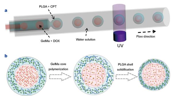
Figure 1
(a) Schematic diagram of a capillary microfluidic system for generating the W/O/W double emulsion templates with polymerized cores; (b) schematic diagram of the fabrication process of the drug loaded GelMa-PLGA core-shell microparticles.
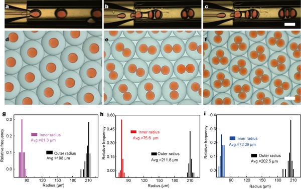
Figure 2
(a–c) Real-time microscopic images of the microfluidic generation process of the W/O/W double emulsion templates encapsulated with tunable number of cores. The scale bar is 100 μm; (d–f) optical microscope image of the monodisperse core-shell double emulsions with one, two and three cores, respectively. The scale bar is 200 μm; (g–i) the size distributions of the inner radiuses and outer radiuses of the double emulsions with one, two and three cores, respectively.
Figure 3
Optical microscopy images (a, e and i) and CLSM images (others) of the DOX and CPT drugs loaded core-shell microparticles. (a–d) Microparticle with single core; (e–h) microparticle with two cores; (i–l) microparticle with three cores. The red and blue fluorescence indicates DOX and CPT, respectively. The scale bar is 100 μm.
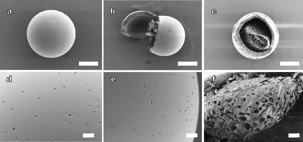
Figure 4
SEM images of the core-shell structure microparticles. (a) External view of a whole microparticle; (b) external view of a shell opened microparticle encapsulated with one core; (c) cross section image of the GelMa-PLGA core-shell microparticle; (d and e) magnified images of shell surfaces of the microparticles in (a, b); (f) magnified image of a partial structure of GelMa core. The scale bars are 100 μm in (a–c) and 10 μm in (d–f).
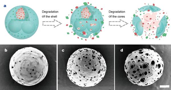
Figure 5
(a) Schematic diagram of the microparticle degradation and its drug release; (b–d) SEM images of the GelMa-PLGA core-shell microparticles during the degradation and release periods of 72, 168, 360 h, respectively. The scale bar is 50 μm.
Figure 6
Figure 7
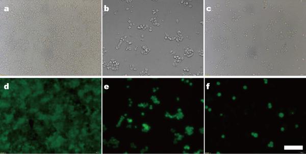
Optical and fluorescence microscopy images of HCT116 cells treated with unloaded microparticles (a, d), only CPT-loaded microparticles (b, e), and DOX-CPT-co-loaded microparticles (c, f) for 24 h, respectively. The scale bar is 50 μm.
Figure 8
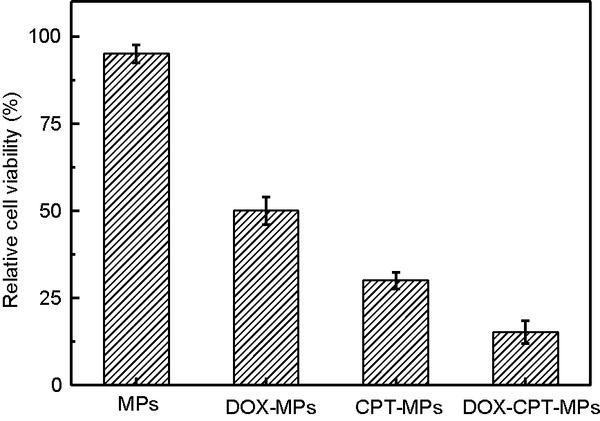
Result of the MTT assay of the HCT116 cells treated with unloaded microparticles (MPs), only DOX-loaded microparticles (DOX-MPs), only CPT-loaded microparticles (CPT-MPs), and DOX-CPT-co-loaded microparticles (DOX-CPT-MPs) for 24 h. Error bars represent standard deviations.
Shell thickness (μm) |
Encapsulation efficiency of CPT (%) |
Encapsulation efficiency of DOX (%) |
Loading content (%) |
22 |
45.80±2.02 |
85.13±0.99 |
4.06±0.02 |
40 |
57.27±0.89 |
89.22±1.12 |
6.17±0.15 |
60 |
60.50±1.24 |
92.73±2.57 |
6.88±0.24 |


Copyright 2019 Science China Press Co., Ltd. 科学大众杂志社有限责任公司 版权所有
京ICP备18024590号-1

 Download PDF

