

Gene therapy targeted to vascular cells represents a promising
approach for prevention and treatment of pathological conditions such
as intimal hyperplasia, in-stent and post-angioplasty restenosis.
In this context, polymeric non-viral gene delivery systems are a safe
alternative to viral vectors but a further improvement in efficiency
and cytocompatibility is needed to improve their clinical success.
Herein, a library of 24 branched polyethylenimine (
The study was supported by the Natural Science and Engineering Research Council of Canada
the Canadian Institute for Health Research
and the Fonds de Recherche du Quebec sur les Natures et Technologies.
Pezzoli D and Tsekoura EK were awarded a post-doctoral and doctoral
scholarship, respectively, from the NSERC CREATE Program in Regenerative
Medicine,
Bahadur KCR and Uluda? H hold ownership position in RJH Biosciences Inc. intended to commercialise the described polymers.
Pezzoli D, Uluda? H, Mantovani D and Candiani G conceived the idea and designed the experiments; Pezzoli D, Tsekoura EK and Bahadur KC R performed the experiments; Pezzoli D and Uluda? H analysed the data and wrote the manuscript with support from Candiani G and Mantovani D. All authors contributed to the general discussion.
Supplementary data are available in the online version of the paper.
[1] Mozaffarian D, Benjamin EJ, Go AS, et al. Executive summary: heart disease and stroke statistics—2015 update: a report from the American Heart Association. Circulation, 2015, 131: 434-441 CrossRef Google Scholar
[2] Shan L, Saxena A, McMahon R, et al. Coronary artery bypass graft surgery in the elderly: a review of postoperative quality of life. Circulation, 2013, 128: 2333-2343 CrossRef PubMed Google Scholar
[3] Marx SO, Totary-Jain H, Marks AR. Vascular smooth muscle cell proliferation in restenosis. Circ-Cardiovasc Interv, 2011, 4: 104-111 CrossRef PubMed Google Scholar
[4] Bauters C, Isner J. The biology of restenosis. Prog Cardiovasc Dis, 1997, 40: 107-116 CrossRef Google Scholar
[5] Arnold JD, Mountain DJH, Freeman MB, et al. Smooth muscle cell polymeric transfection is an efficient alternative to traditional methods of experimental gene therapy. J Surg Res, 2012, 177: 178-184 CrossRef PubMed Google Scholar
[6] Fang YL, Chen XG, Godbey WT. Gene delivery in tissue engineering and regenerative medicine. J Biomed Mater Res, 2015, 103: 1679-1699 CrossRef PubMed Google Scholar
[7] Dean DA. Nonviral gene transfer to skeletal, smooth, and cardiac muscle in living animals. AJP-Cell Physiol, 2005, 289: C233-C245 CrossRef PubMed Google Scholar
[8] Pezzoli D, Chiesa R, De Nardo L, et al. We still have a long way to go to effectively deliver genes!. J Appl Biomater Funct Mater, 2012, 2: 82-91 CrossRef PubMed Google Scholar
[9] Pezzoli D, Candiani G. Non-viral gene delivery strategies for gene therapy: a “ménage à trois” among nucleic acids, materials, and the biological environment. J Nanopart Res, 2013, 15: 1523 CrossRef Google Scholar
[10] Akinc A, Thomas M, Klibanov AM, et al. Exploring polyethylenimine-mediated DNA transfection and the proton sponge hypothesis. J Gene Med, 2005, 7: 657-663 CrossRef PubMed Google Scholar
[11] Mintzer MA, Simanek EE. Nonviral vectors for gene delivery. Chem Rev, 2009, 109: 259-302 CrossRef PubMed Google Scholar
[12] Pezzoli D, Olimpieri F, Malloggi C, et al. Chitosan-graft-branched polyethylenimine copolymers: influence of degree of grafting on transfection behavior. PLoS ONE, 2012, 7: e34711 CrossRef PubMed ADS Google Scholar
[13] Alshamsan A, Haddadi A, Incani V, et al. Formulation and delivery of siRNA by oleic acid and stearic acid modified polyethylenimine. Mol Pharm, 2009, 6: 121-133 CrossRef PubMed Google Scholar
[14] Han S, Mahato RI, Kim SW. Water-soluble lipopolymer for gene delivery. Bioconjugate Chem, 2001, 12: 337-345 CrossRef Google Scholar
[15] Thomas M, Klibanov AM. Enhancing polyethylenimine's delivery of plasmid DNA into mammalian cells. Proc Natl Acad Sci USA, 2002, 99: 14640-14645 CrossRef PubMed ADS Google Scholar
[16]
Zheng
M,
Zhong
Y,
Meng
F, et al.
Lipoic acid modified low molecular weight polyethylenimine mediates nontoxic and highly potent
[17] Neamnark A, Suwantong O, Bahadur RKC, et al. Aliphatic lipid substitution on 2 kDa polyethylenimine improves plasmid delivery and transgene expression. Mol Pharm, 2009, 6: 1798-1815 CrossRef PubMed Google Scholar
[18] Bahadur KCR, Landry B, Aliabadi HM, et al. Lipid substitution on low molecular weight (0.6–2.0kDa) polyethylenimine leads to a higher zeta potential of plasmid DNA and enhances transgene expression. Acta Biomater, 2011, 7: 2209-2217 CrossRef PubMed Google Scholar
[19] Thapa B, Plianwong S, Remant Bahadur KC, et al. Small hydrophobe substitution on polyethylenimine for plasmid DNA delivery: optimal substitution is critical for effective delivery. Acta Biomater, 2016, 33: 213-224 CrossRef PubMed Google Scholar
[20] D'Andrea C, Pezzoli D, Malloggi C, et al. The study of polyplex formation and stability by time-resolved fluorescence spectroscopy of SYBR Green I-stained DNA. Photochem Photobiol Sci, 2014, 13: 1680-1689 CrossRef PubMed Google Scholar
[21] Pezzoli D, Kajaste-Rudnitski A, Chiesa R, et al. Lipid-based nanoparticles as nonviral gene delivery vectors. In: Bergese P, Hamad-Schifferli K (eds.). Nanomaterial Interfaces in Biology: Methods and Protocols, Methods in Molecular Biology. New York: Humana Press, 2013, 1025: 269–279. Google Scholar
[22] Chang KH, Park JM, Lee MY. Feasibility of simultaneous measurement of cytosolic calcium and hydrogen peroxide in vascular smooth muscle cells. BMB Rep, 2013, 46: 600-605 CrossRef Google Scholar
[23] Fan Z, Chen D, Deng CX. Improving ultrasound gene transfection efficiency by controlling ultrasound excitation of microbubbles. J Control Release, 2013, 170: 401-413 CrossRef PubMed Google Scholar
[24] Gresch O, Altrogge L. Transfection of difficult-to-transfect primary mammalian cells. In: Hartley JL (ed.). Protein Expression in Mammalian Cells: Methods and Protocols, Methods in Molecular Biology. New York: Humana Press, 2012, 801: 65–74. Google Scholar
[25] Hsu CYM, Hendzel M, Uluda? H. Improved transfection efficiency of an aliphatic lipid substituted 2 kDa polyethylenimine is attributed to enhanced nuclear association and uptake in rat bone marrow stromal cell. J Gene Med, 2011, 13: 46-59 CrossRef PubMed Google Scholar
[26]
Pezzoli
D,
Giupponi
E,
Mantovani
D, et al.
Size matters for
[27] Suzuki Y, Yeung AC, Ikeno F. The representative porcine model for human cardiovascular disease. J Biomed Biotech, 2011, 2011: 1-10 CrossRef PubMed Google Scholar
[28] Malloggi C, Pezzoli D, Magagnin L, et al. Comparative evaluation and optimization of off-the-shelf cationic polymers for gene delivery purposes. Polym Chem, 2015, 6: 6325-6339 CrossRef Google Scholar
[29] Dube B, Rose L, Sawant K, et al. Cholic acid modified 2 kDa polyethylenimine as efficient transfection agent. Biotechnol Prog, 2013, 29: 1337-1341 CrossRef PubMed Google Scholar
[30] Zhang QF, Luan CR, Yin DX, et al. Amino acid-modified polyethylenimines with enhanced gene delivery efficiency and biocompatibility. Polymers, 2015, 7: 2316-2331 CrossRef Google Scholar
[31] Pezzoli D, Tarsini P, Melone L, et al. RGD-derivatized PEI-PEG copolymers: influence of the degree of substitution on the targeting behavior. J Drug Deliver Sci Tech, 2017, 37: 115-122 CrossRef Google Scholar
[32] Breunig M, Lungwitz U, Liebl R, et al. Breaking up the correlation between efficacy and toxicity for nonviral gene delivery. Proc Natl Acad Sci USA, 2007, 104: 14454-14459 CrossRef PubMed ADS Google Scholar
[33] Aliabadi HM, Landry B, Bahadur RK, et al. Impact of lipid substitution on assembly and delivery of siRNA by cationic polymers. Macromol Biosci, 2011, 11: 662-672 CrossRef PubMed Google Scholar
[34] Hsu CYM, Uluda? H. A simple and rapid nonviral approach to efficiently transfect primary tissue-derived cells using polyethylenimine. Nat Protoc, 2012, 7: 935-945 CrossRef PubMed Google Scholar
[35] Fishbein I, Chorny M, Adamo RF, et al. Endovascular gene delivery from a stent platform: gene- eluting stents. Angiol, 2013, 1: 1000109 CrossRef PubMed Google Scholar
[36]
Saurer
EM,
Yamanouchi
D,
Liu
B, et al.
Delivery of plasmid DNA to vascular tissue
[37] Riessen R, Rahimizadeh H, Blessing E, et al. Arterial gene transfer using pure DNA applied directly to a hydrogel-coated angioplasty balloon. Hum Gene Ther, 1993, 4: 749-758 CrossRef PubMed Google Scholar
[38] Sharif F, Hynes SO, Cooney R, et al. Gene-eluting stents: adenovirus-mediated delivery of eNOS to the blood vessel wall accelerates re-endothelialization and inhibits restenosis. Mol Therapy, 2008, 16: 1674-1680 CrossRef PubMed Google Scholar
[39] Patel SD, Waltham M, Wadoodi A, et al. The role of endothelial cells and their progenitors in intimal hyperplasia. Therapeutic Adv Cardiovascular Disease, 2010, 4: 129-141 CrossRef PubMed Google Scholar
Scheme 1
Synthesis of hydrophobe-substituted
Figure 1
pDNA complexation ability of
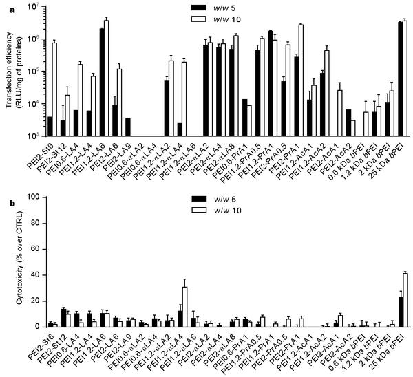
Figure 2
(a) Transfection efficiency and (b) cytotoxicity of
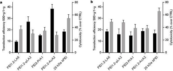
Figure 3
(a) Transfection efficiency and (b) cytotoxicity of
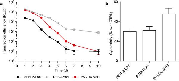
Figure 4
Effect of centrifugation on transfection by
Figure 5
Kinetics of transfection efficiency of selected
Figure 6
(a) Kinetics of transfection efficiency of selected
Polymer |
Substitute |
Feed ratio (mol/mol) |
Degree of substitution (mol/mol) |
BC50 |
PEI2-St6 |
Stearic acid |
6.0 |
2.14 |
0.678 |
PEI2-St12 |
Stearic acid |
12.0 |
4.53 |
8.088 |
PEI0.6-LA4 |
Linoleic acid |
4.0 |
1.09 |
0.364 |
PEI1.2-LA4 |
Linoleic acid |
4.0 |
1.84 |
0.761 |
PEI1.2-LA6 |
Linoleic acid |
6.0 |
2.55 |
0.686 |
PEI2-LA6 |
Linoleic acid |
6.0 |
2.31 |
0.785 |
PEI2-LA9 |
Linoleic acid |
9.0 |
3.20 |
3.609 |
PEI0.6-αLA2 |
α-linoleic acid |
2.0 |
0.80 |
0.320 |
PEI0.6-αLA4 |
α-linoleic acid |
4.0 |
2.30 |
1.269 |
PEI1.2-αLA2 |
α-linoleic acid |
2.0 |
0.94 |
0.289 |
PEI1.2-αLA4 |
α-linoleic acid |
4.0 |
2.45 |
0.297 |
PEI1.2-αLA6 |
α-linoleic acid |
6.0 |
3.17 |
0.578 |
PEI2-αLA2 |
α-linoleic acid |
2.0 |
1.37 |
0.681 |
PEI2-αLA4 |
α-linoleic acid |
4.0 |
2.72 |
1.015 |
PEI2-αLA8 |
α-linoleic acid |
8.0 |
3.68 |
3.899 |
PEI0.6-PrA1 |
Propionic acid |
1.0 |
0.62 |
0.298 |
PEI1.2-PrA0.5 |
Propionic acid |
0.5 |
0.28 |
0.316 |
PEI1.2-PrA1 |
Propionic acid |
1.0 |
0.76 |
0.310 |
PEI2-PrA0.5 |
Propionic acid |
0.5 |
0.15 |
0.304 |
PEI2-PrA1 |
Propionic acid |
1.0 |
0.53 |
0.367 |
PEI1.2-AcA1 |
Acrylic acid |
1.0 |
0.65 |
0.343 |
PEI1.2-AcA2 |
Acrylic acid |
2.0 |
1.21 |
0.430 |
PEI2-AcA1 |
Acrylic acid |
1.0 |
0.51 |
0.355 |
PEI2-AcA2 |
Acrylic acid |
2.0 |
0.86 |
0.643 |
0.6 kDa |
/ |
/ |
/ |
0.278 |
1.2 kDa |
/ |
/ |
/ |
0.215 |
2 kDa |
/ |
/ |
/ |
0.213 |
25 kDa |
/ |
/ |
/ |
0.274 |
?Polymer |
St. Dev. |
PDI |
St. Dev. PDI |
St. Dev. |
|||
PEI1.2-LA6 |
5 |
101 |
16 |
0.30 |
0.11 |
-1.4 |
1.8 |
10 |
135 |
53 |
0.40 |
0.15 |
15.3 |
4.0 |
|
PEI1.2-αLA2 |
5 |
97 |
25 |
0.33 |
0.09 |
21.0 |
1.5 |
10 |
229 |
10 |
0.54 |
0.01 |
23.1 |
0.8 |
|
PEI2-PrA1 |
5 |
98 |
3 |
0.31 |
0.02 |
32.2 |
0.6 |
10 |
112 |
53 |
0.41 |
0.23 |
27.5 |
3.7 |
|
PEI1.2-AcA2 |
5 |
104 |
3 |
0.36 |
0.04 |
26.0 |
6.1 |
10 |
94 |
7 |
0.21 |
0.12 |
22.8 |
4.5 |
|
0.6 kDa |
5 |
1381 |
281 |
1.00 |
0.00 |
14.4 |
5.2 |
10 |
1885 |
1715 |
0.87 |
0.13 |
15.6 |
0.2 |
|
1.2 kDa |
5 |
139 |
7 |
0.45 |
0.03 |
28.9 |
0.6 |
10 |
88 |
0 |
0.03 |
0.01 |
14.9 |
2.1 |
|
2 kDa |
5 |
115 |
13 |
0.27 |
0.05 |
29.1 |
5.4 |
10 |
179 |
152 |
0.39 |
0.23 |
22.0 |
8.0 |
|
25 kDa |
5 |
112 |
20 |
0.35 |
0.05 |
32.8 |
0.7 |
10 |
104 |
16 |
0.29 |
0.09 |
28.2 |
0.9 |


Copyright 2019 Science China Press Co., Ltd. 科学大众杂志社有限责任公司 版权所有
京ICP备18024590号-1

 Download PDF


